. 2016 Sep 26;8(2):1105–1115. doi: 10.1039/c6sc02925f

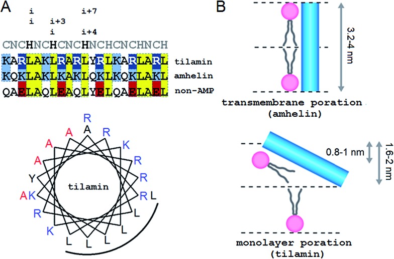
Fig. 1. Tilted antimicrobial helix (tilamin): design and proposed mechanism of membrane insertion. (A, top) Peptide sequences aligned with helical repeats, CNCHNCH, in which hydrophobic residues are separated at i, i + 3 and i, i + 4 helical spacings. Cationic lysines and arginines are in blue and dark blue, respectively, anionic residues in red and absolute identities in yellow. (A, bottom) Tilamin sequence configured onto an α-helical wheel with 3.6 residues per turn. Cationic residues are in blue and the hydrophobic face is underlined. Alanines deuterated for GALA scans are in red. Note: the helical wheel does not reflect the exact spread of the side chains. (B) Schematics of pore edges showing depths of insertion for proposed monolayer and transmembrane poration mechanisms. For clarity, only one peptide (blue cylinder) and one phospholipid per layer are shown (aliphatic chains in grey, headgroups in pink).

Fig. 1