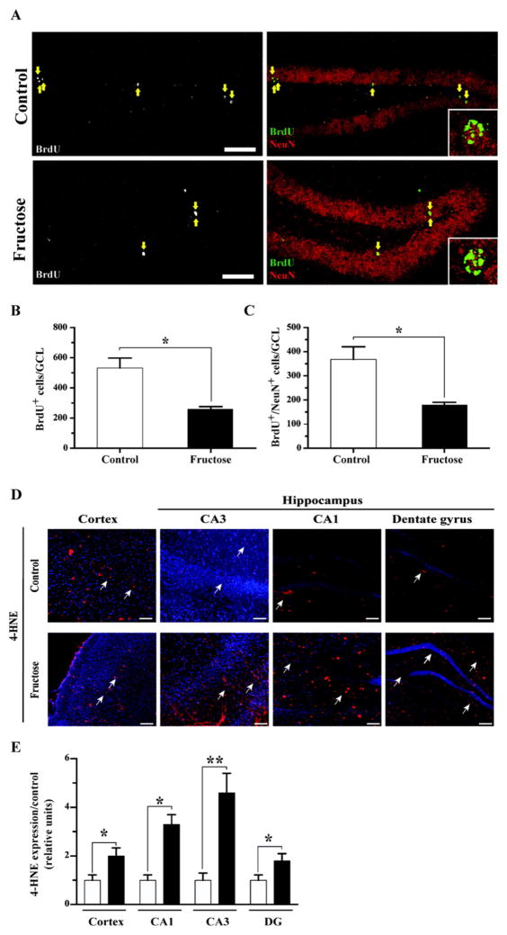
Fig. 5.
MetS reduces the generation of new neurons in the adult hippocampus and increase the levels of oxidative stress. (A) Representative immunofluorescence staining for BrdU and NeuN. The inset shows a higher magnification of BrdU-positive cells that are also positive for NeuN. The arrows indicate BrdU+ cells. Scale bar: 50 μm (original magnification 20×). (B–C) The total number of BrdU+ and BrdU+/NeuN+ cells in the GCL was determined. (D–E) Quantification of the immunofluorescence signal normalized to that under the control conditions. 4-HNE, red; nuclear stain Hoechst, blue. The values are expressed as the means ± SEM of n animals per group. *p < 0.05 and **p < 0.01 based on ANOVA (one-way) followed by Bonferroni post hoc analysis. Scale bar: 100 μm.

