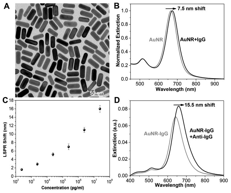
Figure 2.

A) Transmission electron microscopy (TEM) image of AuNRs used as plasmonic nanotransducers. The dimension of the AuNRs is 48×18 nm. B) Extinction spectra showing the LSPR shift after conjugation of AuNR with IgG in solution. The λmax redshifts by 7.5 nm. C) LSPR shift of AuNR-IgG on glass substrate upon exposure to various concentrations of anti-IgG solutions showing the monotonic increase in the LSPR shift with concentration. Error bars represent standard deviations from three different samples. D) Extinction spectra of AuNR-IgG conjugates on the glass substrate before and after exposure to anti-IgG (24 μg mL−1). The λmax redshifts by 15.5 nm.
