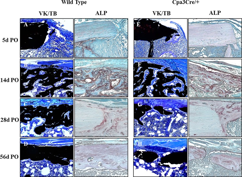
Fig 5. Histochemical analysis of bone in defect/medulla.
Representative images of 5 μm sections of un-decalcified bone stained with von Kossa and toluidine blue (A-H) were compared with the equivalent region of 5μm sections of decalcified bone stained with alkaline phosphatase (ALP). Prominent osteoblasts against osteoid seams can be seen at 14d (F) and 28d (G) PO in Cpa3Cre/+ bones compared with WT bones (B-C). ALP activity peaks at 14 days PO in both WT (B) and Cpa3Cre/+ (F) bones and declines thereafter. Images are representative of N = 7 WT and N = 6 Cpa3Cre/+ at 5d PO; N = 16 WT and N = 11 Cpa3Cre/+ at 14d PO; N = 8 WT and N = 10 Cpa3Cre/+ at 28d PO and N = 6 WT and N = 8 Cpa3Cre/+ at 56d PO.

