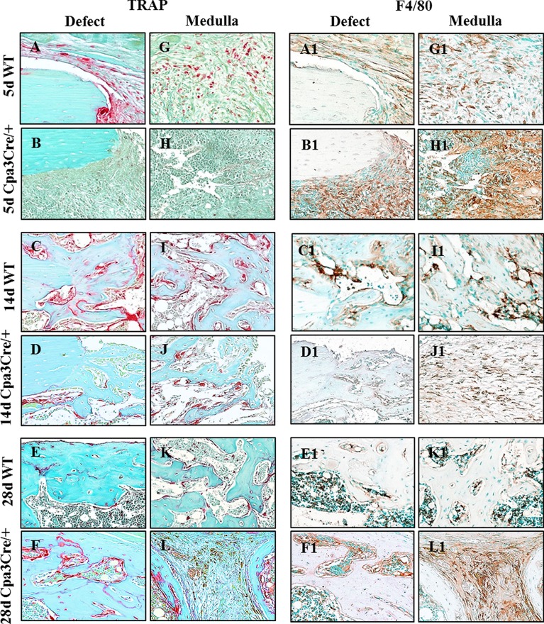
Fig 7. Identification of osteoclasts and macrophages in regenerating bone.
5 μm sections of decalcified bone were stained with tartrate resistant acid phosphatase (TRAP) or immunochemically with the macrophage marker F4/80. Representative images show more TRAP activity in WT than in Cpa3Cre/+ bones at 5d (A vs B) and 14d (C vs D) PO, and less at 28d (E vs F) PO. F4/80 positive macrophages were seen in condensed mesenchyme filling the defect/medulla at 5d PO in both WT (A1) and Cpa3Cre/+ (B1) bones. In WT bone, F4/80 positive cells can be seen lining vessels at 14d (C1) PO and scattered throughout bone marrow at 28d (E1) PO, whereas they were embedded in fibrous tissue in Cpa3Cre/+ bone (F1). Images are representative of N = 7 WT and N = 6 Cpa3Cre/+ at 5d PO; N = 16 WT and N = 11 Cpa3Cre/+ at 14d PO; N = 8 WT and N = 10 Cpa3Cre/+ at 28d PO and N = 6 WT and N = 8 Cpa3Cre/+ at 56d PO.

