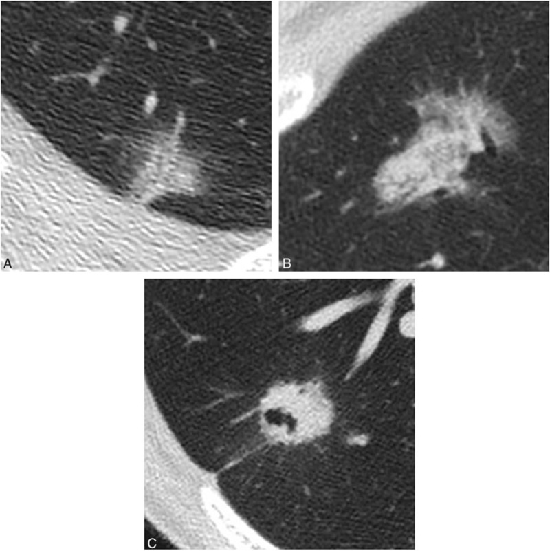
Figure 4.

Each thin-section CT finding in 2 cases of adenocarcinoma in situ (AIS) and in 1 case of invasive adenocarcinoma (IVA). This nodule was histopathologically confirmed as AIS. CT image showed ground-glass nodule, which consisted of 2 kinds of ground-glass densities (Ga + Gc). (B) This nodule was histopathologically confirmed as AIS. CT image showed part-solid nodule, in which solid portion included air bronchiologram without any disruptions and irregular dilatations. Solid portion correlated to the pathological collapse area. (C) This nodule was histopathologically confirmed as IVA with acinar, papillary, and micropapillary cells. CT image showed irregular solid nodule including air bronchiologram with disruptions and irregular dilatations. Pleural indentation can be seen.
