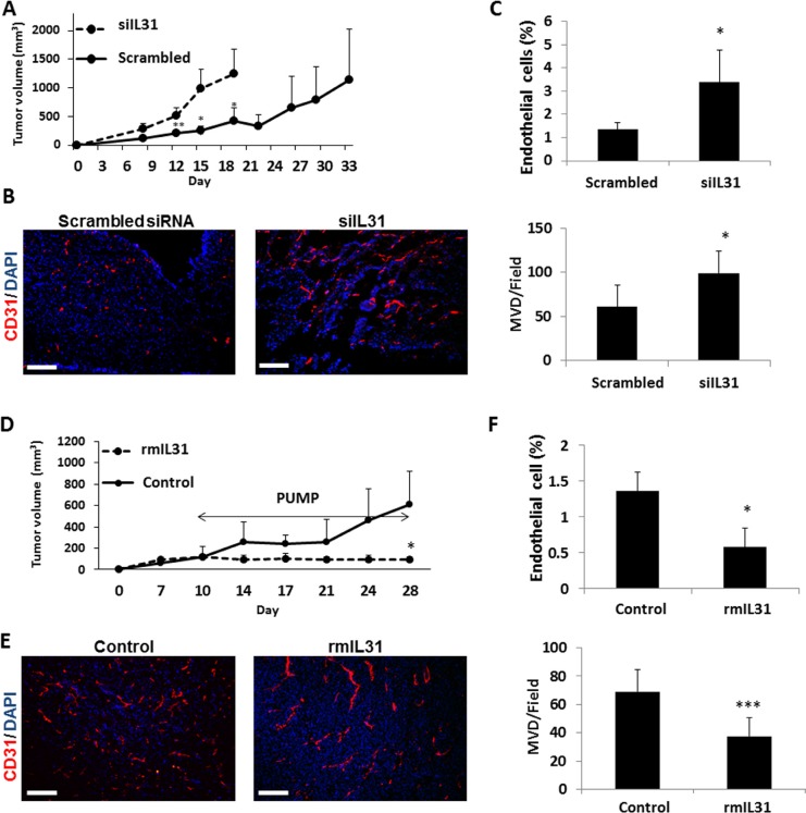
Figure 3. IL31 inhibits tumor growth and angiogenesis.
(A–C) Control or IL31 siRNA MC38 cells (1 × 10⁶) were subcutaneously implanted into the flanks of 8-week old C57Bl/6 mice (n = 4–5 mice/group). Tumor growth was assessed with a caliper using the formula width2 × length × 0.5 (A). At end point, tumors were removed and divided into two equal parts. One part was sectioned (B) and the other part was prepared as a single cell suspension (C). Tumor sections were immunostained for CD31, an endothelial cell marker (red). Nuclei were stained with DAPI (blue). Scale bar = 200 μm (B). Tumor single cell suspensions were assessed for the percentage of endothelial cells by flow cytometry (C). (D–F) MC38 cells (1 × 10⁶) were implanted into the flanks of 8-week old C57Bl/6 mice (n = 4–5 mice/group). Tumors were allowed to grow until they reached a size of 100 mm3, at which point mice were implanted with micro-osmotic pumps containing rmIL31 (administered at a dose of 0.7 μg/day) or PBS. Tumor growth was assessed regularly (D). At end point, tumors were removed and divided into two equal parts. One part was sectioned for endothelial cell staining (E) and the other part was prepared as a single cell suspension for the evaluation of endothelial cell percentage by flow cytometry (F), as in (B–C). **, 0.01 > p > 0.001; ***p < 0.001.

