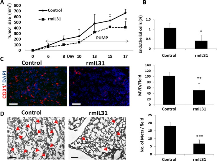
Figure 4. IL31 inhibits angiogenesis and lung metastasis in 4T1 metastatic breast carcinoma.
4T1 cells (0.5 × 10⁶) were implanted into the mammary fat pad of 8 week old BALB/c mice (n = 5 mice/group). After 3 days, mice were implanted with micro-osmotic pumps containing rmIL31 (administered at a dose of 0.7 μg/day) or PBS (control). Tumor growth was assessed regularly (A). At end point, tumors were removed and divided into two equal parts. One part was sectioned (B) and the other part was prepared as a single cell suspension (C). Tumor sections were immunostained for CD31, an endothelial cell marker (red). Nuclei were stained with DAPI (blue). Scale bar = 200μm (B). Tumor single cell suspensions were assessed for the percentage of endothelial cells using flow cytometry (C). (D) Lungs were harvested at end point. Left panel: Lung sections were stained with H&E to visualize metastatic lesions (red arrows). Representative images are shown. Scale bar = 200 μm. Right panel: Metastatic lesions per field were quantified (n > 15 fields/group) *p < 0.05; **, 0.01 > p > 0.001; ***p < 0.001.

