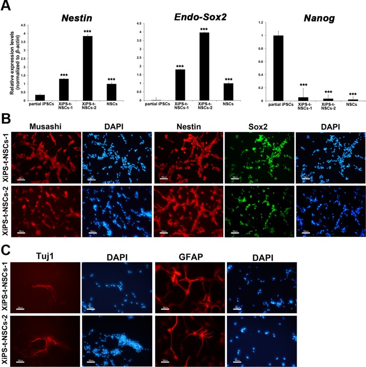
Figure 3. Characterization of in vivo NSCs from teratomas derived from partially reprogrammed cells.
(A) Real-time RT-PCR analysis for endo-Sox2 (endogenous), Nestin, and Nanog in partial iPSCs, XiPS-t-NSCs, and brain-derived NSCs. Relative expression levels were calculated by normalizing to β-actin; data represent mean ± SEM. Student’s t-test: ***p < 0.001 (B) Immunofluorescence staining of NSC markers Musashi, Nestin, and Sox2 in XiPS-t-NSCs, with DAPI counterstaining of nuclei; scale bar = 50 μm. (C) XiPS-t-NSCs differentiated into neurons (Tuj1+) and glial cells (GFAP+) in vitro. Nuclei were counterstained with DAPI; scale bar = 50 μm.

