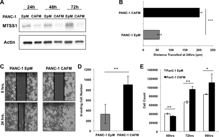
Figure 4. Treatment with CAF media results in decreased MTSS1 and increased proliferation and migration in PANC-1 cells.
(A) Western blot analysis of PANC-1 cells treated with either epithelial-conditioned media (EpM) or CAF-conditioned media (CAFM) and harvested at various timepoints. (B) PANC-1 cells were treated with either EpM or CAFM and submitted to a scratch assay for 24 hours. (C) Representative images of PANC-1 cells treated with either EpM or CAFM during scratch assay analysis. Images were taken at 10× magnification. (D) PANC-1 cells were plated for a transwell migration assay with either EpM or CAFM in the upper chamber. After 48 hours, cells were fixed to the membrane, stained with hematoxylin, and counted for analysis. (E) PANC-1 cells were incubated in either CAFM or EpM and harvested at various timepoints to observe any changes in proliferative ability. *p-value < 0.05, **p-value < 0.001, ***p-value < 0.0001.

