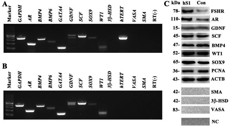
Figure 2. Phenotypic feature of the immortalized human Sertoli cells.
(A–B) RT-PCR showed the expression of AR, BMP4, BMP6, GATA4, GDNF, SCF, SOX9, WT1, hTERT, 3β-HSD, SMA, and VASA in the immortalized human Sertoli cells (A) and primary human Sertoli cell (B). GAPDH was used as a loading control of total RNA, and RNA sample without RT (RT-) but with PCR of GAPDH primers served as a negative control. (C) Western blot revealed the proteins of FSHR, AR, GDNF, SCF, BMP4, WT1, SOX9, PCNA, 3β-HSD, VASA, and SMA in the immortalized human Sertoli cells (hS1) and primary Sertoli cells (Con). ACTB was used as a loading control of proteins, while replacement of primary antibodies with PBS served as negative controls (NC).

