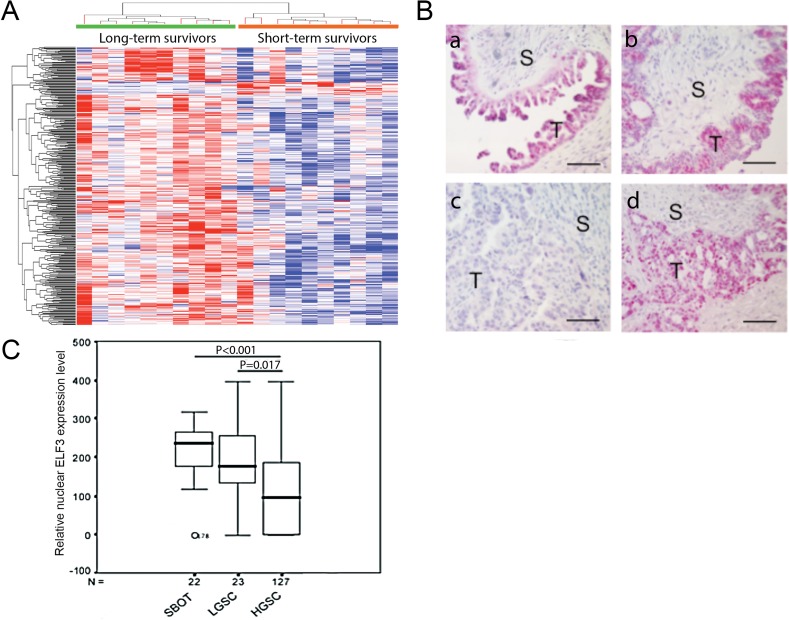
Figure 1. ELF3 expression in ovarian tumor tissue samples.
(A) Heat map showing that ELF3 was identified as one of the upregulated transcription factors in ovarian cancer cells according to transcriptome profiling analysis. (B) Immunolocalization of nuclear ELF3 in (a) SBOT, (b) LGSC, and (c-d) HGSC samples. S, stroma; T, tumor tissue. Bar = 50 μm. (C) Box plot showing nuclear ELF3 expression in SBOT, LGSC, and HGSC samples. The 25th percentile is shown at the bottom of the box, the 75th percentile is shown at the top, and the whiskers represent 95% confidence intervals.

