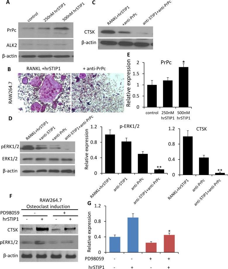
Figure 8. Activation of STIP1-PrPc-ERK1/2 signaling in osteoclast differentiation.
(A) Expression of PrPc and ALK2 in the total cell lysate from hrSTIP1 treated-RAW264.7 cells. (B) TRAP staining of osteoclasts induced in RAW264.7 cell line upon treatment with hrSTIP1 (500 nM) or hrSTIP1+anti-PrPc (10 μg/ml) for 48 hours. (C) Expression of CTSK in the total cell lysate from indicated treatment in RAW264.7 cells. (D) Expression of p-ERK1/2 and total ERK1/2 in the total cell lysate from indicated treatment in RAW264.7 cells. (E) Quantification of the western blot analysis (A, C, D) with three repeats. Expression of PrPc and CTSK were normalized to the level of β-actin, and expression of pERK1/2 was normalized to total ERK1/2. *p < 0.05, vs control; **p < 0.01, vs RANKL+hrATIP1. (F) Suppression the activation of endogenous ERK1/2 signaling by pre-treatment with 15 μM PD98059 for 2-h inhibited the hrSTIP1-induced CTSK protein expression in RAW264.7 cells. (G) Quantification of the western blot analysis of CTSK expression with three repeats. *p < 0.05, vs PD98059 -/hrSTIP1+.

