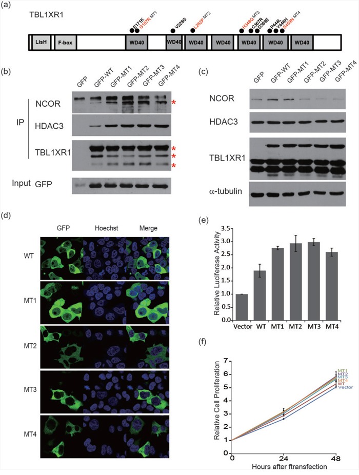
Figure 5. Functional characterization of TBL1XR1 WD40 domain alternations.

a. Schematic diagram of the structure of TBL1XR1. Mutants occurred in the WD40 domain as indicated with circles. b. Co-immunoprecipitation of TBL1XR1 mutants. TBL1XR1 physically interacts with NCoR repressor complexes. Mutant TBL1XR1 interacts with NCoR and HDAC3 more frequently than wild-type TBL1XR1. c. Expression of NCoR protein levels in cells transfected with wild-type or mutant TBL1XR1. d. Fluorescence images of TBL1XR1 and Hoechst staining and merged images in 293T cells transfected with each TBL1XR1 mutant. e. NF-κB activity assay measuring the effects of each TBL1XR1 mutant. f. Cell proliferation comparison of TBL1XR1 mutants expressed in cells over 48 h using a CCK-8 assay.
