
Figure 6. Tspan8 silencing reduces ILK activity with a concomitant decrease in β1 integrin-dependent adhesion and clustering.
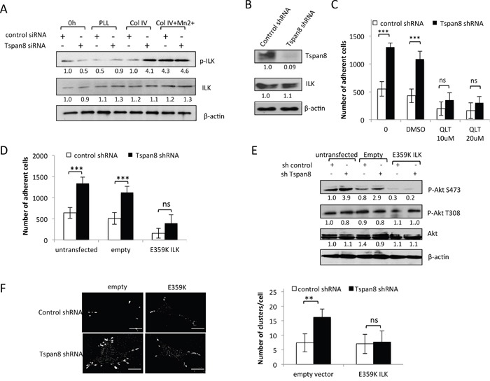
A. T1C3 cells silenced or not for Tspan8 were plated on poly-L-lysine (PLL) or collagen IV (col IV) with or without Mn2+ and allowed to adhere for 2 h. Cell lysates before (0 h) or after adherence were analyzed by Western blot with total or phosphorylated ILK mAbs (representative blot of 3 independent experiments). The band intensities were normalized to β-actin signal. B. Western blot analysis of Tspan8 and ILK in invasive T1C3 cells stably transduced with control or Tspan8 shRNAs expressing plasmids. β-actin was used as a loading control. Of note, Tspan8 knockdown had no effect on the ILK protein expression levels. C. These cells were pre-treated or not with QLT0267 at 10 μM or 20 μM or vehicle (DMSO), plated on collagen IV-coated plates and subjected to adhesion assay. Bars represent the mean number of adherent cells ± SD from a representative experiment (n=2, each in sixplicate). ***, p < 0.001, Student t test. D-F. T1C3 cells stably silenced for Tspan8 were transfected or not with empty or ILK-E359K plasmids, plated onto collagen IV-coated plates and subjected to adhesion assays (D) western blot analysis (E) and microscopy (F). (D) Bars represent the number of cells that adhere to collagen IV-coated plates (mean ± SD of sixplicate samples from one representative experiment; n=3). (E) Representative western blot of total (Akt) and phosphorylated (P-Akt) Akt (n=3). β-actin was used as a loading control and a internal reference for band quantification. (F). T1C3 cells plated onto collagen IV-coated coverslips were stained with 12G10 mAb. Left panel, representative confocal microscopy images (n=2; scale bar: 10 μm). Right panel, mean number of β1 integrin clusters per cell ± S.D. from up to 40 cells over 2 separate experiments. **, p < 0.05, Student t test.