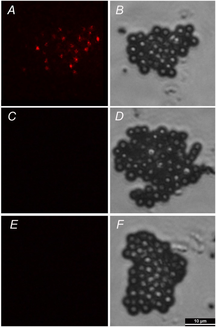
Fig 2. SCNT embryos-derived exosomes were identified by immunofluorescence.
The exosomes were bound to beads of a size that was in the detection range of the fluorescence microscope (4-μm diameter latex beads). The beads were then bound to fluorescence-conjugated antibody against CD9. Images were taken under epifluorescence (A, C, E) and DIC (B, D, F). A,B: embryos-derived exosomes; C,D: IgG negative control; E,F: embryo-free culture medium control. Bar, 10 μm.

