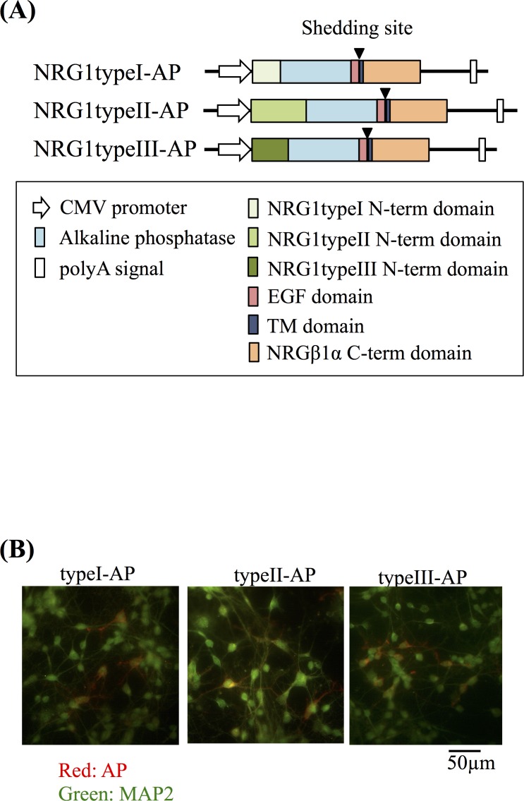
Fig 4. Gene structures of expression vectors encoding AP-tagged types I–III NRG1 precursors.
(A) Diagram of the pRc-CMV/NRG1-AP expression vectors. The sequences encoding the human placental AP tag (blue), NRG1 type I–III-specific N-terminal domains (orange, type I; red, type II; yellow, type III). The EGF (light green) and transmembrane domains (deep green) are marked. (B) Following transfection of cells with individual expression vectors, AP immunoreactivity on the surface of living cells was visualized together with MAP2. Live cortical neurons expressing AP-tagged NRG1 type I-III precursors were treated with the anti-AP antibody to visualize NRG1 precursors on the cell surface. After fixation, cells were incubated with a MAP2 antibody followed by fluorescent secondary antibodies (red for AP and green for MAP2). Scale bar = 50 μm.

