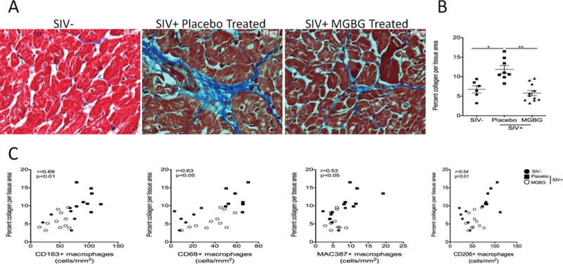
Figure 3. MGBG treatment prevents an increase in cardiac fibrosis in cardiac tissues similar to levels seen in uninfected controls.

(A) Collagen, a marker of fibrosis, in cardiac tissues was measured by staining cardiac tissue with a trichrome stain. ImageJ Analysis software was used to determine the percent collagen (blue dye) per total tissue area from 20 200× non-overlapping fields of view with the average percent collagen per total tissue area calculated for each animal. (B) There was a significant 2.05-fold decrease in the percent collage per total tissue area when comparing placebo controls to MGBG treated animals. There was no significant difference between the percent collagen per total tissue area between uninfected and MGBG treated animals. Statistical analysis was performed using analysis of variance (ANOVA). If the ANOVA was significant (p<0.05) between two groups, a posthoc non-parametric Mann-Whitney t-test was performed. (C) Using Spearman rank correlation analysis, we found positive correlations between increasing numbers of CD163+ (r=0.69), CD68+ (r=0.63), MAC387+ (r=0.53) and CD206+ (r=0.54) macrophages and increasing percent collagen per total tissue area. * p<0.05, ** p<0.01. Scale bar equals 20 μm.
