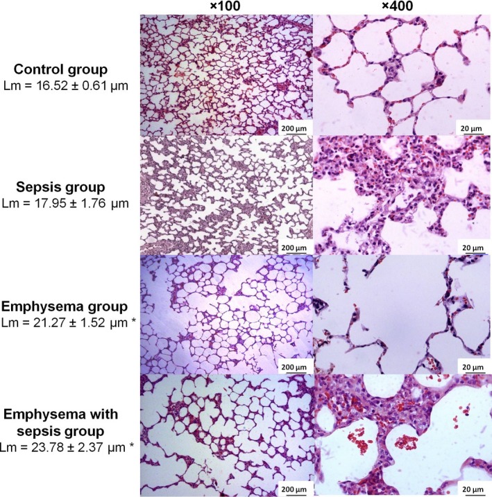
Figure 2.

Photomicrographs of lung parenchyma stained with haematoxylin–eosin. Note that sepsis group showed alveolar wall thickening, neutrophils in the interstitium and in the airspace, and proteinaceous debris in the airspace. Emphysema group showed airspace enlargement and alveolar wall destruction. Emphysema with sepsis showed emphysematous abnormalities (airspace enlargement and alveolar wall destruction) combined to acute lung injury findings (alveolar wall thickening, neutrophils in the interstitium and in the airspace, and proteinaceous debris in the airspace). The mean linear intercept (Lm) values are shown as means and standard deviation. Statistical analysis was performed using one‐way anova followed by Tukey test. *P < 0.05 compared with control and sepsis groups.
