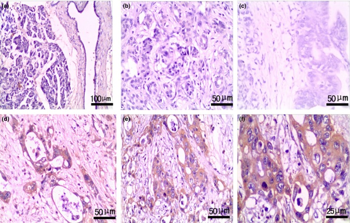
Figure 2.

Expression of Ihh in pancreas with benign and malignant lesions. EnVision™ immunohistochemistry: (a) negative Ihh expression in pancreatic normal tissues (×100); (b) negative Ihh expression in chronic pancreatitis tissues (×200); (c) negative Ihh expression in PIN II tissues (×200); (d) positive Ihh expression in well‐differentiated PDA (×200), tumour size of 3 cm and TNM stage I; (e) positive Ihh expression in poorly differentiated PDA (×200), tumour size of 4 cm and TNM stage IV; (f) magnified image from e, Ihh staining was localized in the cytoplasm of epithelial cells in poorly differentiated PDA (×400).
