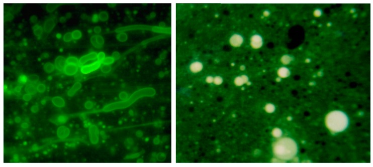
Figure 5.
Membranous vesicles self-assemble from mixtures of fatty acids and alcohols such as decanoic acid and 1-decanol [61]. The vesicles shown here (left panel) were stained with rhodamine 6G and photographed by fluorescence microscopy (400× original magnification) [68]. Typical vesicles shown in the micrograph are ~1–10 μm in diameter. After a single dehydration cycle (right panel), the vesicles readily encapsulated macromolecules such as ~600 nt duplex DNA. The DNA was stained with acridine orange, an intercalating fluorescent dye.

