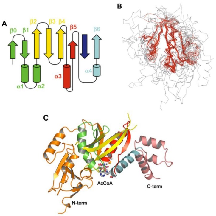
Figure 9.
(A) Topology of the GNAT fold. From the N-terminus, secondary structural elements are colored green (β1, α1, α2), yellow (β2–4), red (α3, β5) and blue (α4, β6). The dark green (β0) N-terminal strand is not completely conserved and the deep blue C-terminal strand may be from the same monomer, or contributed by another; (B) Superposition of 15 GNAT structures. Residues in which the RMSD is <2.7 Å are highlighted in red; (C) Yeast Hat1 histone acetyltransferase in complex with Ac-S-CoA (1BOB.pdb). Reproduced with permission from ref. [94].

