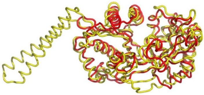
Figure 10.
Superposition of FemX (red) with S. aureus FemA (yellow, PDB 1LRZ). The overall structures are similar: RMSD is 2.8 Å for the 309 Cα atoms). The major structural difference is the absence in FemX of the coiled-coil region constituted by two helices (residues 246–307; found in SerRS) inserted in FemA domain 2. Reproduced with permission from ref. [97].

