FIG 1.
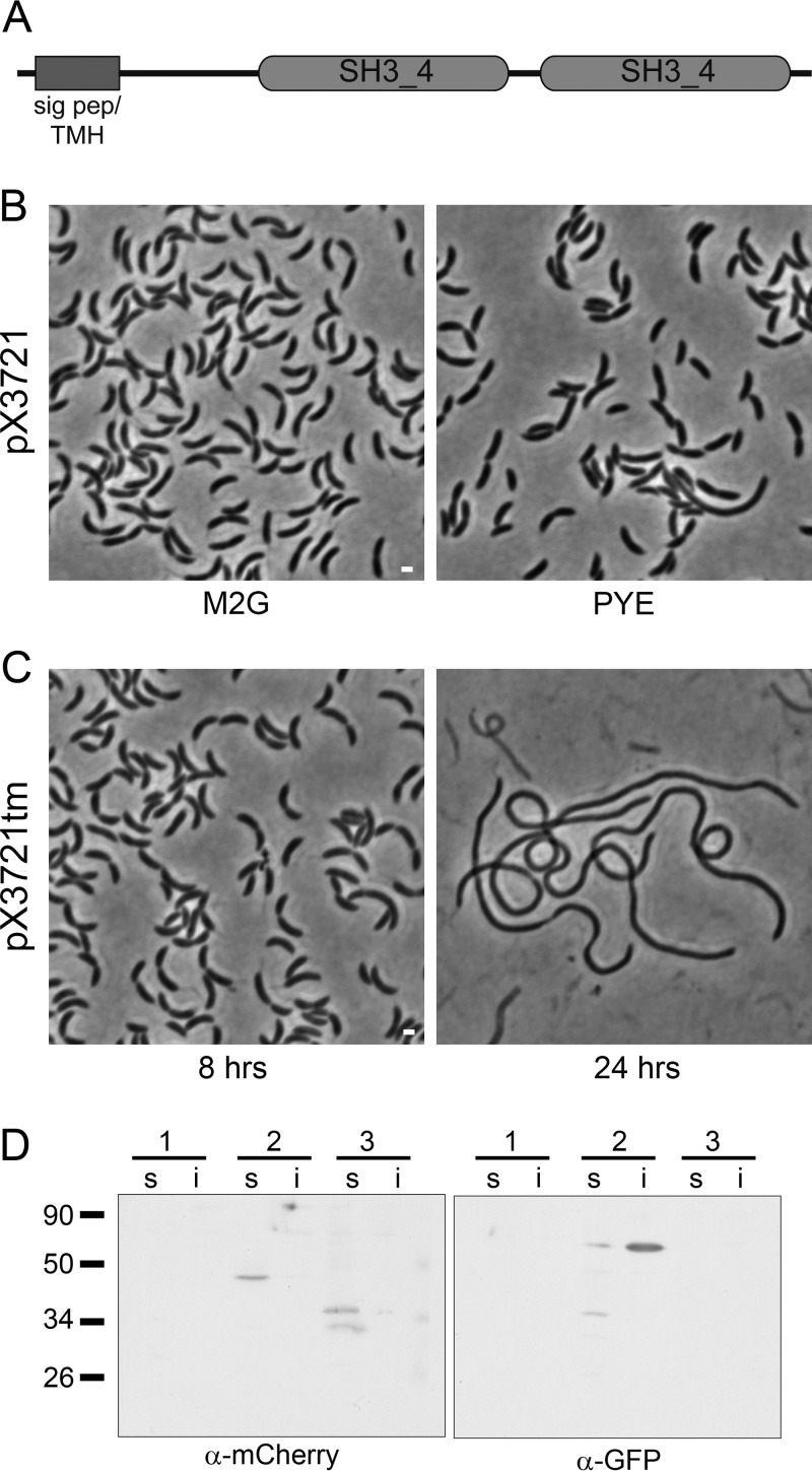
Depletion of DipI causes cell filamentation. (A) The domain structure of DipI is shown. (B) A strain (SP2) with a single chromosomal copy of dipI under the control of a xylose-inducible promoter (pX3721) was grown in minimal medium (M2G) or rich medium (PYE) in the absence of xylose. (C) A strain (SP3) with a single chromosomal copy of dipI fused with the sequence for the degradation signal encoded by tmRNA under the control of a xylose-inducible promoter (pX3721tm) was grown from an overnight culture in M2G medium without xylose for 8 or 24 h. (D) Presence of DipI-mCherry in the soluble fraction. Western blots of soluble (s) and insoluble (i) fractions of the following strains were probed with the indicated antibodies: 1, CB15N (wild type); 2, SP22 (expressing DipI-mCherry and Venus-FtsN protein fusions); and 3, CJW2959 (expressing periplasmic mCherry). Expected molecular masses of the proteins in kilodaltons were as follows: periplasmic mCherry, 32.8; Venus-FtsN, 55; and periplasmic DipI-mCherry, 44.2. Bars, 1 μm.

