FIG 2.

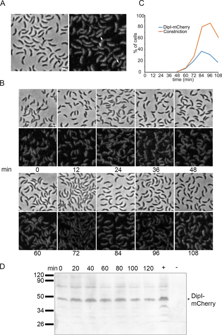
DipI is recruited late to the division site. (A) Localization of DipI-mCherry in an unsynchronized cell population. Cells from a culture of strain SP15 grown in minimal medium (M2G) were observed when the culture reached an OD660 of 0.3. Arrows indicate cells with no visible constriction in which DipI-mCherry was already localized at midcell. (B) Time-lapse images for localization of DipI-mCherry in a synchronized cell population. A culture with an OD660 of 0.3 was synchronized, and aliquots were taken for observation every 12 min. Empty arrows indicate cells with deep constriction that did not show localization of DipI. (C) Quantification of the localization of DipI to the division site. The percentage of cells that showed constriction or localization of DipI was determined in 300 cells for each time point after synchronization. (D) Presence of DipI-mCherry during the cell cycle. Total cell extracts from a synchronized culture of the SP15 strain were obtained every 15 min and the presence of DipI-mCherry was determined by Western blotting. Total cell extracts from unsynchronized SP15 and CB15N cultures were used as positive and negative controls, respectively. Migration of molecular weight markers is shown at the left. The asterisk indicates the migration of the DipI-mCherry protein. Bars, 1 μm.
