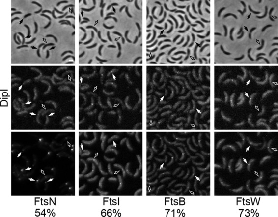
FIG 3.

DipI is recruited late to the division site. The colocalization of DipI-mCherry with other inducible cell division protein fusions was quantified in exponential cultures. Top panels, phase-contrast images; middle panels, DipI-mCherry fluorescence images; bottom panels, fluorescence images of Venus fusions with the division protein indicated at the bottom of the image (strains from left to right: SP22, SP26, SP21, and SP23). Empty arrows indicate dividing cells where localization of DipI-mCherry was expected but not observed; solid arrows indicate division sites or cell poles were colocalization was observed. The percentages of colocalization are shown at the bottom. Percentages were calculated only from cells that showed localization of the indicated division protein at the division site (n ≈ 300). Bar, 1 μm.
