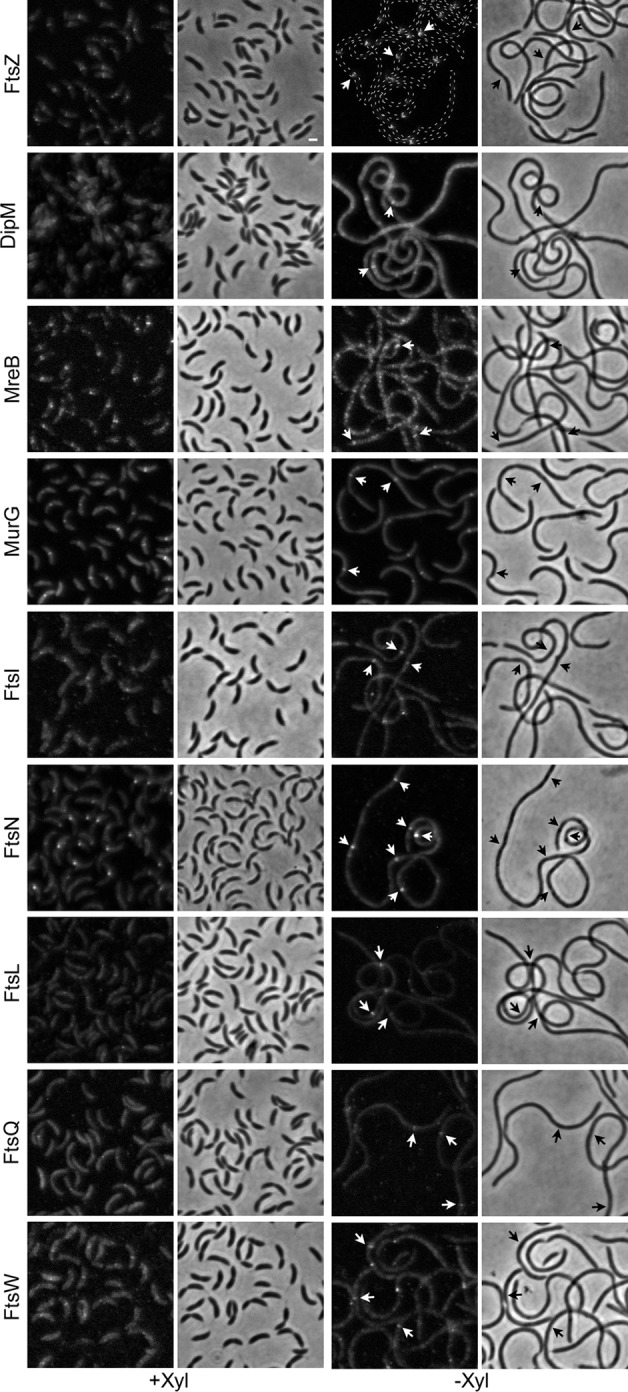
FIG 4.

Mature divisomes form in the absence of DipI. The localization of different cell division proteins was determined in cells expressing DipI (+Xyl) or depleted of DipI (−Xyl). Fluorescent protein fusions of the different cell division proteins (indicated at the left of each row) were introduced as second copies and expressed from a vanillic acid-inducible promoter. The murG gene was substituted with the allele coding for the fluorescent fusion. All the strains were grown in rich medium (PYE) in the presence or absence of xylose. Depletion was carried out as described in Materials and Methods. When required, vanillic acid was added 3 h before observation. Arrows indicate localization of the division proteins in sites where no constriction was visible. From top to bottom, the strains used are as follows: SP4, SP6, SP7, SP8, SP9, SP5, SP27, SP28, and SP10. Bar, 1 μm.
