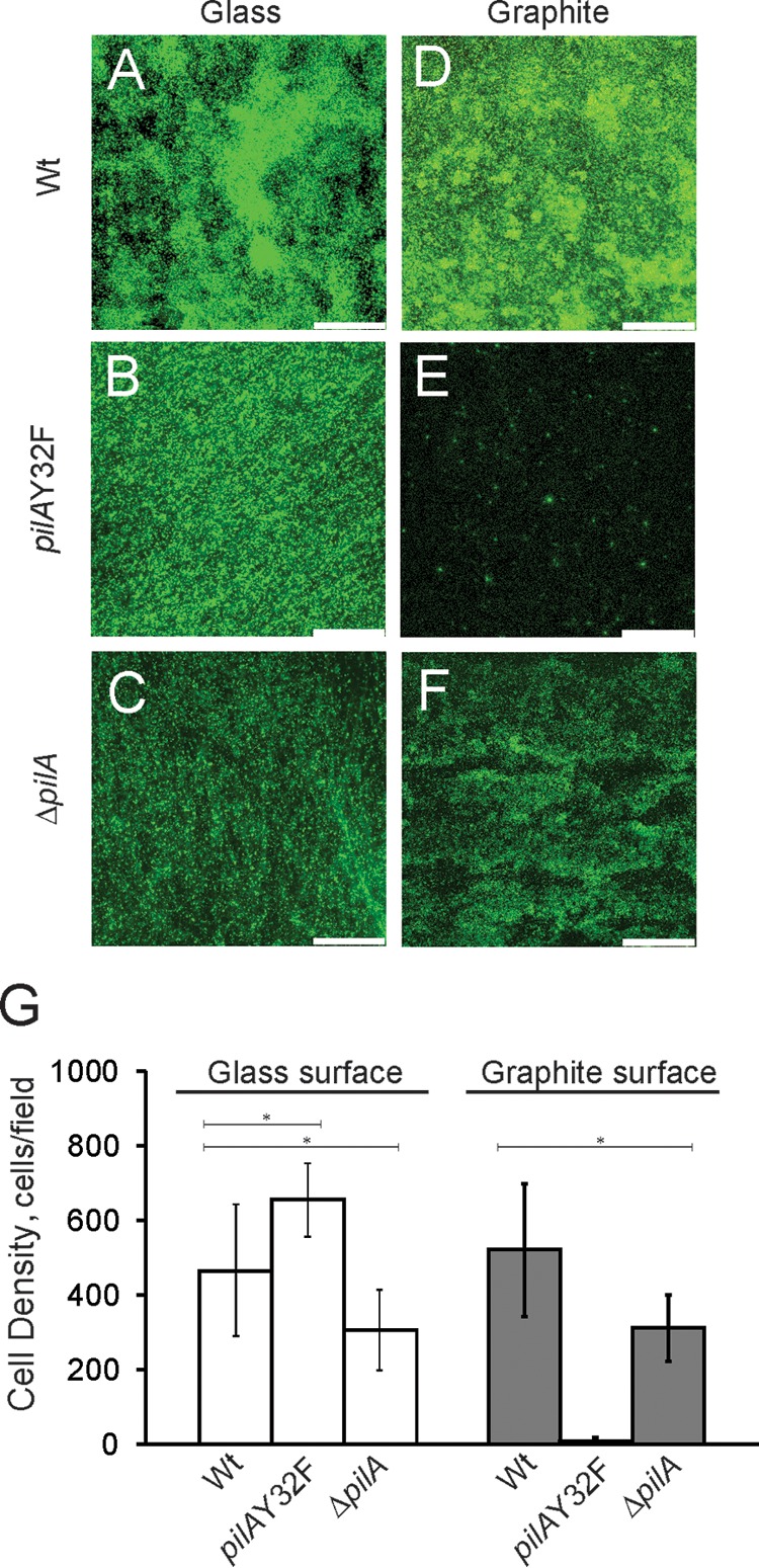
FIG 4.

(A to F) Confocal microscopic images of biofilms formed with the isogenic wild-type DL100, pilAY32F, and ΔpilA in-frame deletion strains on glass and graphite surfaces. Cells were incubated with soluble electron donor and acceptor (10 mM acetate and 40 mM fumarate, respectively) on glass/graphite slips for 4 days under anaerobic conditions. Bars, 75 μm. (G) Average numbers of cells of the wild-type DL100, pilAY32F, and ΔpilA strains attached to glass and graphite surfaces after 4 days of incubation. The results are the averages and standard errors of the means from six biological replicates in two independent experiments. Statistical analysis was performed by Student's t test; *, P < 0.001.
