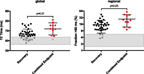
Fig. 1.

Predictive value of global and regional T2 time. Displayed are global T2 values on the left and regional T2 values with respect to area fraction exceeding 80 ms on the right. Patients having experienced MACE or were admitted to hospital due to heart failure are coloured in red. Reference range (mean ± SD) of global and regional T2 values for healthy controls are given as grey bars. Initial global T2 time of patients who experienced endpoint (n = 11) was 71.8 ± 5.7 ms while it was 66.8 ± 4.9 ms in those patients who did not (n = 45) (p = 0.01). In fact, the myocardial fraction with abnormal T2 time at first presentation was larger in patients who reached endpoint (combined endpoint: 27.5 ± 14.9% vs. no endpoint: 15.1 ± 8.7%. p = 0.03). Abbreviations: Fraction >80 ms = Percentage of myocardial fraction with T2 time >80 ms
