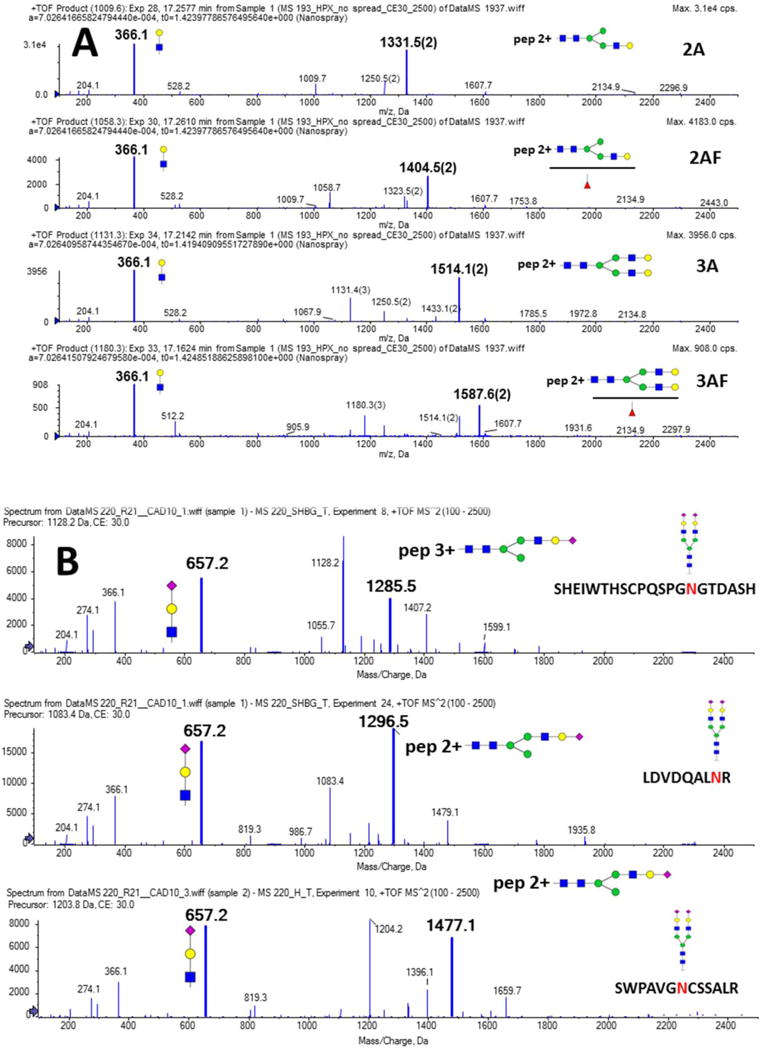
Fig 1.

Soft fragmentation of the following analytes: A. SWPAVGNCSSALR glycopeptide of hemopexin with four different glycan structures attached (2A, 2AF, 3A, and 3AF); B. biantennary sialylated glycan (2A2SA) attached to the peptide backbones of SHBG (SHEIWTHSCPQSPGNGTDASH and LDVDQALNR) and hemopexin (SWPAVGNCSSALR). Structure, m/z, and charge state (M-1) of the major soft fragment (Y-ion) as well as the related oxonium ions (loss of glycan fragment, charge 1) are indicated in the figure.
