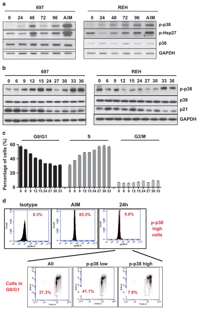
Figure 1.
p38α/β signaling upon ALL proliferation in vitro. (a) 697 cells (left panel) and REH cells (right panel) were cultured in vitro and assayed for p38α/β- and Hsp27-phosphorylation by western blotting at different time points (0, 24, 48, 72 and 96 h, as indicated). Cells treated with 10 μg/ml Anisomycin (AIM) for 15 min were processed in parallel as controls. (b) 697 cells (left panel) and REH cells (right panel) were serum starved for 24 h and then cultured in full serum conditions for the indicated time points. Western blotting was performed for p38α/β, p38α/β-phosphorylation, p21 and glyceraldehyde 3-phosphate dehydrogenase (GAPDH). (c) In parallel to the experiment in b, cells were allowed to incorporate BrdU for 1 h at the indicated time points and stained with 7-AAD and an anti-BrdU antibody. Cell cycle phases were determined by flow cytometry. (d) 697 cells were grown in vitro for 24 h, incubated with BrdU for 1 h and stained with a fluorescein isothiocyanate-labeled p-p38α/β antibody or the respective isotype control. Subsequent staining with 7-AAD was also performed. After fluorescence-activated cell sorting analysis, the bulk of cells (All), p-p38α/β-low and the 10% cells with highest p-p38α/β (p-p38α/β-high) were gated and cell cycle phases were assayed as indicated. Cells in G0/G1 are found in the red gate. 7-AAD, 7-amino-actinomycin D.

