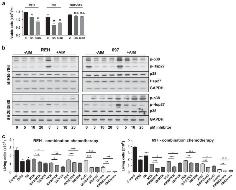
Figure 2.
Cell counts and signaling upon p38α/β inhibition in vitro. The experiments shown are representative experiments of at least two repetitions. (a) REH (left panel), 697 (middle panel) and SUP-B15 cells (right panel) were incubated with 10 μM SB203580 (SB) or BIRB-796 (BIRB) for 72 h. The number of viable cells was counted using standard Trypan blue exclusion. *P<0.05; NS, not significant; t-test. (b) REH (left panel) and 697 cells (right panel) were incubated with ascending concentrations of BIRB-796 (upper panels) or SB203580 (lower panels) for 48 h. Dimethyl sulfoxide (0 μM lanes) was used as a vehicle control. Cells were either directly lysed (− AIM) or stimulated with 10 μg/ml Anisomycin for 15 min before lysis (+AIM). The indicated markers were assayed by western blotting. (c) REH (left panel) and 697 cells (right panel) were incubated with 20 μM BIRB-796 (BIRB) or SB203580 (SB) alone or in combination with chemotherapeutic drugs for 48 h, as indicated. MTX, 30 nM Methotrexate (REH and 697); VCR, 1 nM (REH) and 2 nM (697) Vincristine; DXM, 100 nM (REH) and 50 nM (697) Dexamethasone; AraC, 50 nM Cytarabine (REH and 697); Dauno, 100 nM Daunorubicine (REH and 697). Standard chemotherapeutics were used at concentrations nearest to the IC50 of the individual drug and cell line. Other concentrations are depicted in Supplementary Figure 3. *P <0.05, **P <0.01, ***P <0.001. C, control; GAPDH, glyceraldehyde 3-phosphate dehydrogenase.

