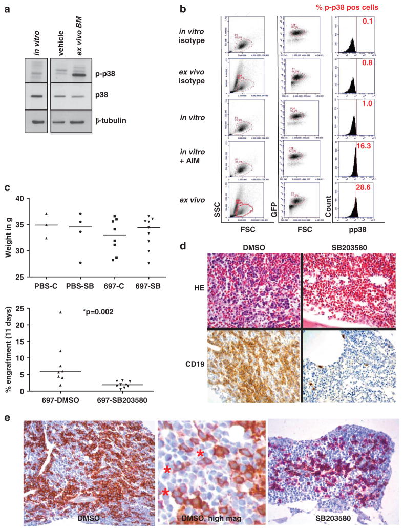
Figure 3.
Induction of p38α/β phosphorylation in 697 cells in the bone marrow of avian xenografts. (a) Cells cultured in vitro (lane 1) for 24 h after CD19 magnetic-activated cell sorting (MACS) and cells recovered from the bone marrow of a turkey embryo after 12 days in vivo (day E23) after CD19 MACS (lane 3) were subjected to western blot analysis for p38α/β phosphorylation and total p38 levels. Bone marrow from a turkey embryo injected with phosphate-buffered saline (lane 2) was processed in parallel as a control. (b) 697-GFP cells were injected into turkey embryos and recovered from the bone marrow on day E23 (panels 2+5). 697-GFP cells were also cultured in vitro for 24 h in parallel (panels 1+3). Cells cultured in vitro for 24 h were induced with 10 μg/ml Anisomycin (AIM) for 15 min (panel 4). Cells from all conditions were stained with a PE-labeled p-p38α/β antibody. After fluorescence-activated cell sorting (FACS) analysis, cells were gated for the lymphoblast gate (P1) and then for GFP-positivity (P2 dependent on P1). Cells in the P2 gate were assayed for p38α/β phosphorylation. The percentage of p-p38α/β-positive cells is depicted in red. (c) 697-GFP cells were xenografted into turkey embryos as in a. Control eggs were injected with PBS. Eggs were treated with 10 mg/kg body weight SB203580 (SB) or 0.1% dimethyl sulfoxide as vehicle control every other day by intra-amniotic injection. Embryos were killed on day E23 by rapid decapitation, weighed and cells were recovered from the bone marrow of one femoral bone and assayed for GFP-positivity by FACS analysis. The upper panel shows the body weight in g in the respective groups. The lower panel shows engraftment as % GFP-positive cells with respect to all nucleated cells in the bone marrow. Nucleated cells in avian systems include erythrocytes. (d) The other femoral bone from c was fixed in formalin solution, embedded in paraffin, cut and stained by standard hematoxylin/eosin (HE) or immunohistochemistry for human CD19 (CD19). (e) Target specificity of p38α/β inhibition assayed by double staining of p-Hsp27 (brown) and CD19 (purple). In DMSO-treated xenografts (left panel), a dark brown staining indicating p-Hsp27 positivity is evident, whereas in SB203580-treated xenografts (right panel), only purple staining is visible. The middle panel represents a digital high-magnification view showing CD19-positive purple cells also (*).

