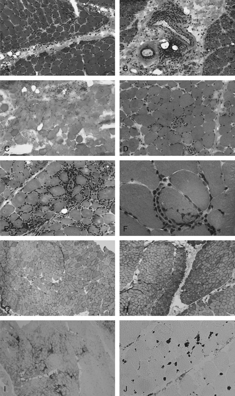
FIGURE 1.

Pathologic features of idiopathic inflammatory myopathy. A–C. Pathologic dermatomyositis: perifascicular atrophy (A), perivascular lymphocytic infiltrates (B), and microinfarct (C). D. Necrotizing myopathy: numerous necrotic and regenerative fibers. E–F. Pathologic aspects suggestive of polymyositis: endomysial infiltrates (E) and invasion of a nonnecrotic muscle fiber by lymphocytes (F). G–I. Examples of MHC-1 overexpression: diffuse (G), diffuse with perifascicular reinforcement (H), focal (I). J. Microthrombi of C5b-9 in intramuscular capillaries. (A–F: Hematoxylin-eosin stain, G–J: immunohistochemistry. A–C original magnification × 100, D–E × 200, F × 630, G × 80, H–I × 40, J × 250.).
