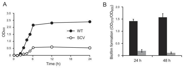
Fig. 2.
Growth curves and biofilm formation in AM3 medium at 37°C. (A) Growth curves of WT and SCV. (B) Biofilm formation by WT (black) and SCV (gray) on microtiter plates after 24-h and 48-h incubations. The amounts of biofilms were normalized by cell densities (OD600). Data are the average of at least six replicate wells and the standard deviations are shown.

