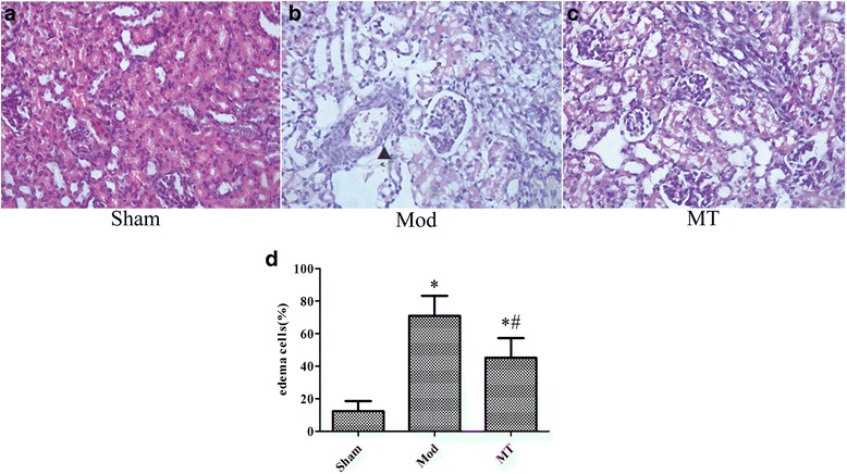
Fig. 3.

Renal cortex under a 400 × light microscope after H-E staining. Similar treatment as Fig. 2 induced significant swelling of the renal tubular epithelial cells and interstitial tissue edema, which was improved by melatonin treatment in H-Estained samples (a and b). However, there were few changes regarding the glomeruli in these groups. In addition, the swollen renal tubular epithelial cells were quantified in the total renal tubular epithelial cells in the indicated groups.↗ represents cytotoxic edema, and▲ represents vasogenic edema
