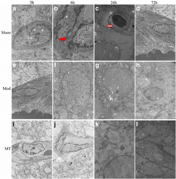
Fig. 5.

Cerebral glial cells and capillaries assessed via a transmission electron microscope. HIBD could result in a remarkable pathologic change at 24 h after injury, including clearly decreased organelles and their extraordinary swelling, fused or disappeared intercellular junctions and additional stenosis and contracture of the capillary lumens (e-h) compared with sham group (a-d). Melatonin treatment could reduce the severity of pathology after injury (i-l). Red solid arrows represent tight junctions, red hollow arrows represent capillary lumens, and ▲ represents cell vacuoles
