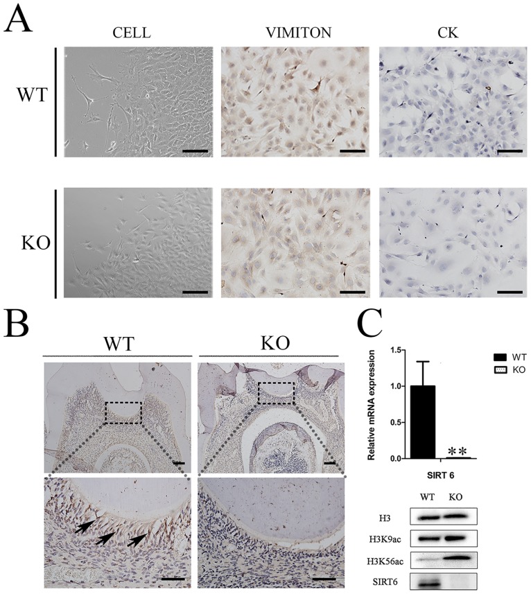
Fig 3. Expression of SIRT6 in mouse dental pulp tissue.
(A) The DMCs were either fusiform or triangular in shape; they were positive for vimentin, and negative for cytokeratin. (B) The higher magnification images of Immunohistochemial (IHC) staining showed that SIRT6 was expressed in the nuclei of mature odontoblasts in the WT group. The KO group in contrast showed SIRT6-negative expressions (bars 50μm). Cell nuclei were visualized with hematoxylin. (C) The mRNA and protein levels of Sirt6 were almost undetectable in the KO mice group. The gene expression level was normalized to GAPDH (n = 6). An assessment ofH3K9ac and H3K56ac in both the WT and KO groups was carried out by western blot analysis. This analysis found that the KO groups experienced a significant increase in these histones. (n≥3) *p<0.05; **p<0.01versus CO. CO = control.

