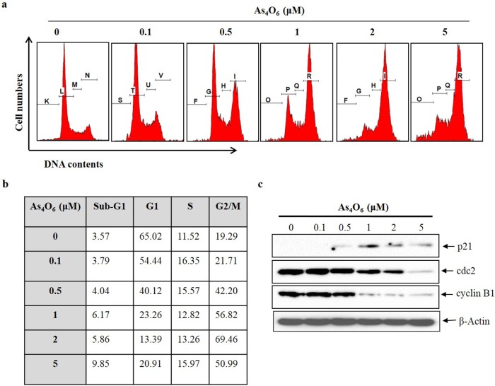
Fig 2. As4O6 induced G2/M cell cycle arrest of SW620 cells.
The cells were seeded at the density of 5x10⁴ cells per ml. (a) To quantify the cell cycle phase distribution, the cells were treated with As4O6 at 0, 0.1, 0.5, 1, 2 and 5 μM concentrations for 24 h and stained with PI followed by flow cytometry was performed. (b) Quantitative data represents G2/M arrest was induced by As4O6 on SW620 cells in a dose dependent manner. (c) The cells were treated with As4O6 at 0, 0.1, 0.5, 1, 2 and 5 μM concentrations for 24 h. Total cell lysates were resolved by SDS-polyacrylamide gels and transferred onto nitrocellulose membranes. The membranes were probed with the p21, cyclin B1, cdc2 antibodies. To confirm equal loading, the blot was stripped of the bound antibody and reprobed with the anti β-actin antibody. The proteins were visualized using an ECL detection system. The data are shown of three independent experiments.

