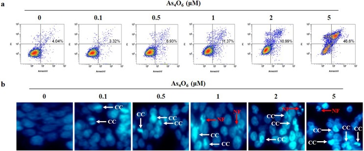
Fig 3. As4O6-induced apoptosis in SW620 cells.
The cells were seeded at the density of 5x10⁴ cells per ml. (a) To quantify the extent of As4O6-induced apoptosis, the cells were treated with As4O6 at 0, 0.1, 0.5, 1, 2 and 5 μM concentrations for 24 h and Annexin V was analyzed by flow cytometry. (b) After fixation, the cells were stained with DAPI solution to observe apoptotic body. Stained nuclei were then observed under fluorescent microscope using a blue filter (Magnification, X 400). CC represents chromatin condensation; NF represents nuclear fragmentation. The data are shown of three independent experiments.

