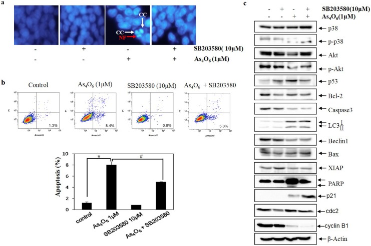
Fig 7. The role of p38 MAPK in As4O6-induced cell death in SW620 cells.
SW620 cells were treated with SB203580 (10 μM) before As4O6 (1 μM) for 48 h. (a) To confirm apoptosis, the cells were stained with DAPI solution after fixation. Stained nuclei were then observed under fluorescent microscope using a blue filter (Magnification, X 400). CC represents chromatin condensation; NF represents nuclear fragmentation. (b) Apoptosis was assessed by Annexin V/PI flow cytometry assay. (c) The cells were lysed and equal amount of the lysate was separated by SDS-polyacrylamide gels, and then transferred to nitrocellulose membranes. The membranes were probed with the indicated antibodies, and detected by an ECL detection system. To confirm equal loading, the blot was stripped of the bound antibody and reprobed with the anti ß-actin antibody. The data are shown as mean ± SD of three independent experiments. * p<0.05between the As4O6-treated and the untreated control group; # p<0.05 compared between the combination treatment group (SB203580 and As4O6) and As4O6 alone treatment group.

