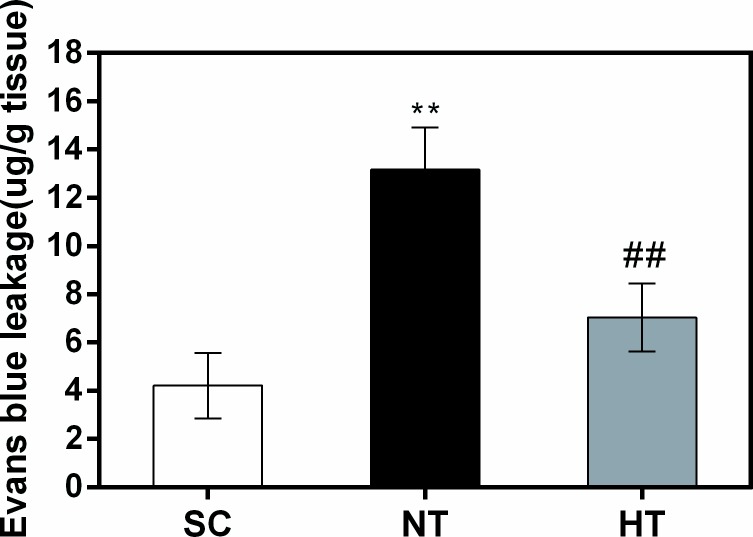
Fig 5. Blood–brain barrier permeability in cortical tissues at 24 hours after CA and resuscitation.
Blood–brain barrier permeability was quantitatively evaluated via leakage of Evans blue in the SC (n = 4), NT (n = 4), and HT (n = 4) groups. Data are presented as means ± sd. SC, surgery control group; NT, non-hypothermia group; HT, mild hypothermia group. **P<0.01 versus SC group; ##P<0.01 versus NT group.

