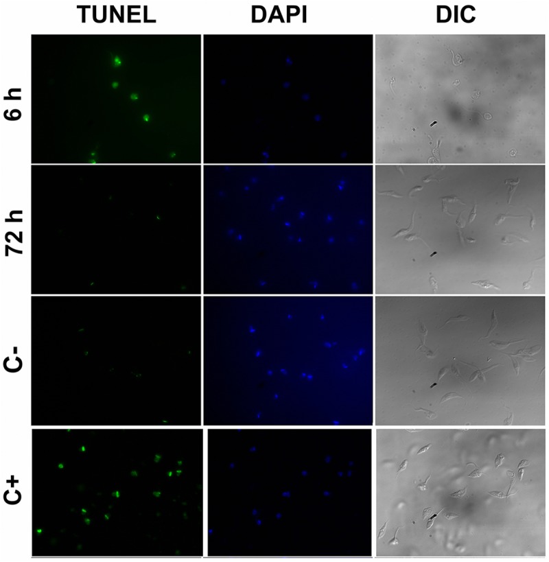
Fig 5. Fluorescence micrographs of TUNEL-stained epimastigotes.

Epimastigote cells were treated with DNAse I (apoptosis positive control, C+), without treatment (negative control, C-) or 200 and 30 μM isotretinoin for 6 and 72 h, respectively. Parasites were fixed and permeabilized after the TUNEL reaction (green) and stained with DAPI (blue). Images acquired by fluorescence microscopy under 100x objective showed positive TUNEL in DNAse I control and cells treated with 200 μM isotretinoin for 6 h. Differential interference contrast (DIC) images showed the differences in cell morphology (partially or fully rounded cells) after 6 h isotretinoin exposure. The TUNEL protocol was detailed under Methods.
