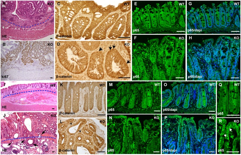
Fig 4. The IEC-specific hnRNPI knockout mice develop colorectal neoplasia with highly active Wnt and NF-κB signaling.
(A) HE–stained section shows non-invasive neoplasia in the colonic epithelium of the knockout mouse. The dotted line indicates the position of muscularis mucosae. (B) Immunohistochemical staining with an anti-Ki67 antibody shows hyperproliferation in colonic neoplasia. (C) and (D) Immunohistochemical staining with an anti-β-Catenin antibody shows nuclear translocation of β-Catenin in the colonic epithelium of the knockout mice (D, arrows). In the wild-type colonic epithelium, β-Catenin is membrane localized (C). (E) to (H) Immunofluorescence staining with an anti-p65 antibody shows p65 nuclear translocation in the colonic epithelium of the knockout mice (F and H). p65 is mainly localized in the cytoplasm in the control mice (E and G). Nuclei were counterstained with DAPI (G and H). (I) and (J) HE–stained section shows invasive adenocarcinomas in the prolapsed rectum of the knockout mouse. The dotted line indicates the position of muscularis mucosae. Note a few glands have invaded through the muscularis mucosae into the submucosa (arrows in J). (K) and (L) Immunohistochemical staining shows nuclear accumulation of β-Catenin in the rectal epithelial cells in the knockout mice (arrowheads in L), whereas in the control rectal epithelium, β-Catenin is cell membrane localized (K). (M) to (R) Immunofluorescence staining shows nuclear translocation of p65 in the rectal epithelium of the knockout mice (N, P and R). The control mice showed cytoplasmic localization of p65 (M, O and Q). Nuclei were counterstained with DAPI (O and P). (Q) and (R) High magnification shows p65 nuclear localization in the rectal epithelial cells. WT, wild-type; KO, knockout. Scale bars, 50 μm.

