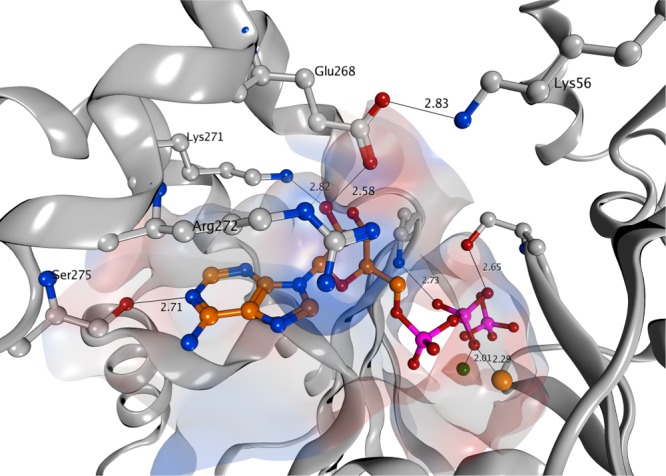
Figure 3.

Crystal Structure of ADP/Pi bound to HSP72. PDB 3ATU, important hydrogen bonding interactions, with their distances in Å, and residues are indicated. The key salt-bridge interaction between lysine-56 and glutamic acid-268 was measured at 2.8 Å (2 SF). Orange and turquoise spheres represent sodium and magnesium ions, respectively. Only selected residues are shown and solvent has been omitted for clarity.
