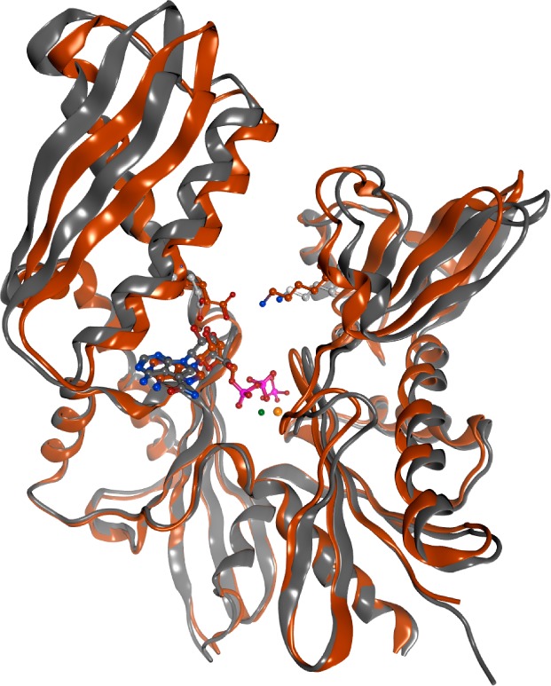
Figure 5.

Induced open and induced closed conformations of HSP72. PDB 3ATU and 5AQZ, the copper-colored structure represents the co-crystal structure of ADP/Pi bound to the HSP72-NBD in the induced closed conformation due to interactions of the phosphate groups through hydrogen bonds with the glycine rich loops and stabilized by the salt-bridge. The gray-colored structure represents the co-crystal structure of sangivamycin 10 bound to the HSP72-NBD. The overlay shows sangivamycin 10 bound to a more open NBD conformation, in which the formation of the Glu-Lys salt-bridge is not possible. Only selected residues are shown and solvent has been omitted for clarity.
