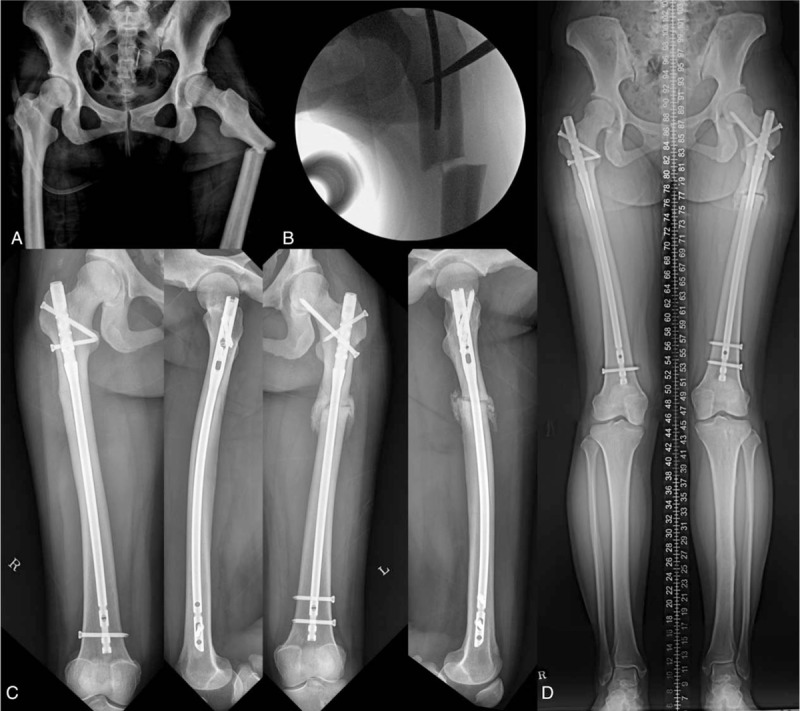
Figure 1.

Atypical femur fracture of the patient. (A) Preoperative radiographs of the patient with pycnodysostosis. (B) Intraoperative C-arm image shows a long osteotome used to open and widen the intramedullary canal. (C) Four months postoperatively, radiographs show that the left femur exhibited substantial bridging callus across the fracture, and the right femur was healed. (D) Scanogram shows good alignment with an incomplete union of the fracture.
