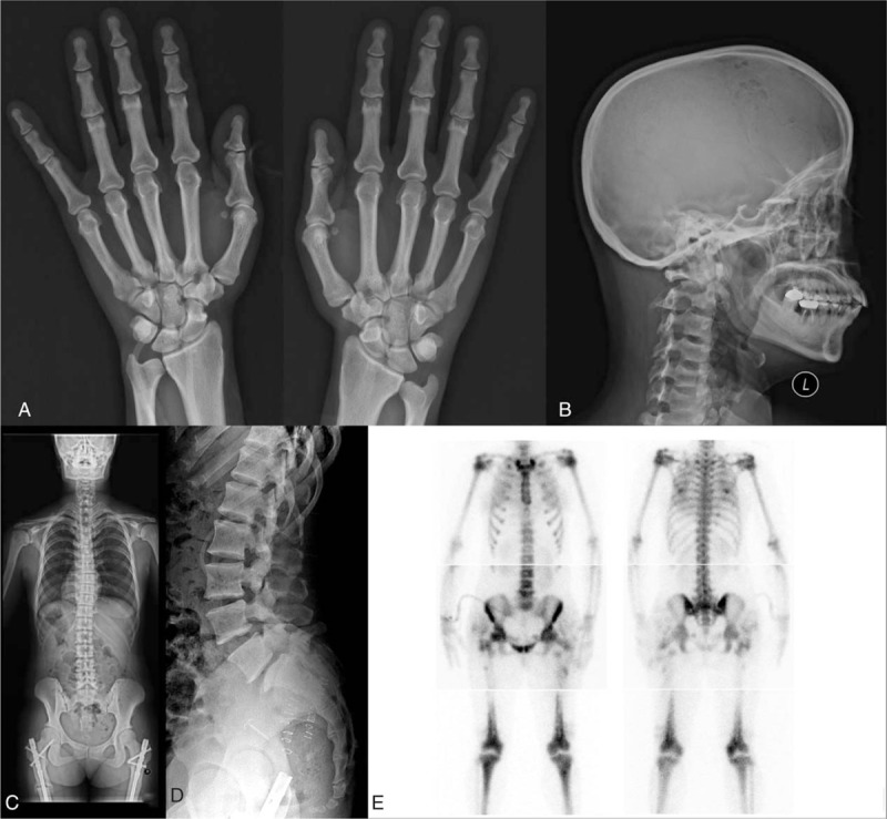
Figure 2.

Skeletal survey of the patient. (A) Acroosteolysis of the distal phalanges was not observed in the hand radiograph. (B) Lateral cranial film shows thickening at the base of the skull and frontal bossing. (C) Mild scoliosis was observed on the whole spine posteroanterior (PA) radiograph. (D) Spondylolysis of the L5 vertebrae was observed on the lateral lumbar spine radiograph. (E) Multiple hot uptake was observed on the whole body bone scan.
