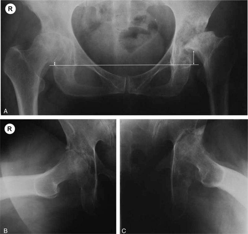
Figure 3.

Bilateral RPOH in a 64 years old female patient—AP view of the pelvis (A), and axial views of the right (B) and left (C) hip. Femoral head ascension was determined on the AP view of the pelvis: the horizontal line connects the radiologic teardrops, while the vertical lines connect the horizontal to the inferior junction of the femoral head and neck. The right hip presents grade II RPOH, while the left hip is grade III: complete disappearance of the joint line, multiple geodes and deforming of the acetabulum and femoral head, major femoral head osteolysis and ascension >0.5 cm above the level of the radiological teardrop.
