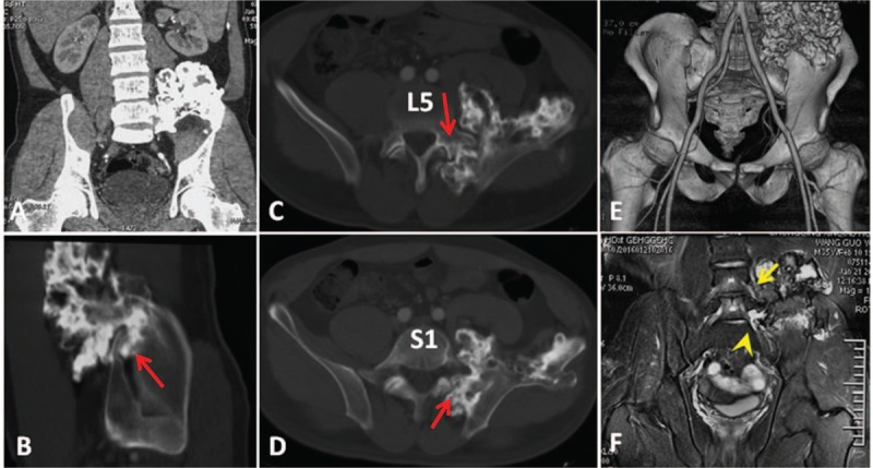
Figure 1.

(A) Preoperative coronal, (B) sagittal, and (C, D) axial CT scans showing that the tumor (red arrow) involves the ilium, left sacroiliac joint, left transverse process, pedicle, and vertebral body of L5 and the sacrum. (E) The anterior aspect of the preoperative 3-dimensional computed tomography scan displays the proximity of the iliac vessels and the tumor. (F) Magnetic resonance image demonstrating that the tumor is adhesive to the L4 (yellow arrow) and L5 (yellow arrow head) nerve roots.
